


Why splints, is not a diagnoses and what shin could be causing my leg pain?
Hello and welcome to my first blog for UKRunChat. Let me introduce myself, my name is Nick Knight, a Musculoskeletal and Sports Podiatrist, based in the south of England with clinics in Southampton, Winchester, Basingstoke and London. I run a website nksportspodiatry.co.uk, which is currently been updated and shall offer advice to runners. I plan to offer monthly blogs to help advise and educate runners and the active person. Each topic will be decided by you.
Personally I am a keen sportsman, playing hockey, squash, along with been a keen cyclist. I am used to compete as a 100m runner, and now enjoy running as a hobby. I concentrate on treating and assessing runners and getting people from injury back to running, also helping try and prevent the injury occurring again. I have treated all types of runners from new comers in couch to 5k, right up to elites at the London Marathon and 2012 Paralympics.
The aim of the blogs is to be easy reading for runners, whilst offering the current evidence, I shall offer my opinion. I enjoy good debate and always open discuss the blogs in more detail. There will be a science nature to these blogs.
The topic of this month’s blog is exercise induced leg pain.
Exercise induced leg pain commonly gets term ‘shin splints’ which is incorrect, don’t panic, there are still many medical professionals and even some journals still use this poor term. Their excuse is that everyone understands the term shin splints, however unless we stop using the term how will anyone learn. So hopefully by the end of this blog we will not be see the term ‘shin splints’ used within UKRunChat or UkTriChat.
There are many causes of exercise induced leg pain some very common and other very rare affecting around 1:100,000 people. Each cause of exercise induced leg pain has their own presentation and symptoms, each requiring the correct treatment. I hope you can begin to see the issue with labelling all the diagnoses with 1 broad term of ‘shin splints’.
To help keep things simple and not too long I shall focus on, 3 of the most common causes of exercise induced leg pain that I see in clinic on a day-to-day basis. I shall go through how they present in clinic and some of the current treatments used.
We shall go through:-
- Medial tibial stress syndrome, this is the main term that people call shin splints.
- Stress fracture.
- Biomechanical overload syndrome
Before we get started I thought it would be a good idea to run through some basic anatomy. The leg consists of 2 bones the tibia, the big shinbone and the fibula the smaller bone on the outside of the leg. The tibia is the main weight-bearing bone. The Fibula has been to be called a coat hanger the muscles. I’m not going to go into detail on the muscles in the leg however the leg is split into 4 compartments the anterior (front), posterior (back), deep posterior (behind the posterior) and lateral (outside).

Medial tibial stress syndrome
If anyone wants to more detailed reading on medial tibial stress syndrome please click here for more detail blog I’ve recently wrote on medial tibial stress syndrome.
Medial tibial stress syndrome is the most common cause of exercise induced leg pain I see in clinic and very common with in runners and jumpers. We are still not entirely sure what medial tibial stress syndrome is, there are a couple of theories.
The 1st theory being the anatomical theory that proposes certain muscles within the leg pull against the periosteum (the tissue surrounding the bone), however there is debate as to which muscle is involved, this is due to anatomical variation, meaning that the muscles in the body to not always present as you see in the textbook.
The 2nd theory is the biomechanical/bone stress theory. This theory suggests that the pain is due to increased bone stress as you apply a compression force to the tibia it bends at its narrowest point which, tends to be the area of where you get the pain.
Just for your information, if you are interested, bone is strongest when you apply a compression force, followed by a tensile force so stretching the bone, then weakest when applying a torsional force i.e. twisting the bone.
(Reilly & Burstein.1975)
How does medial tibial stress syndrome present?
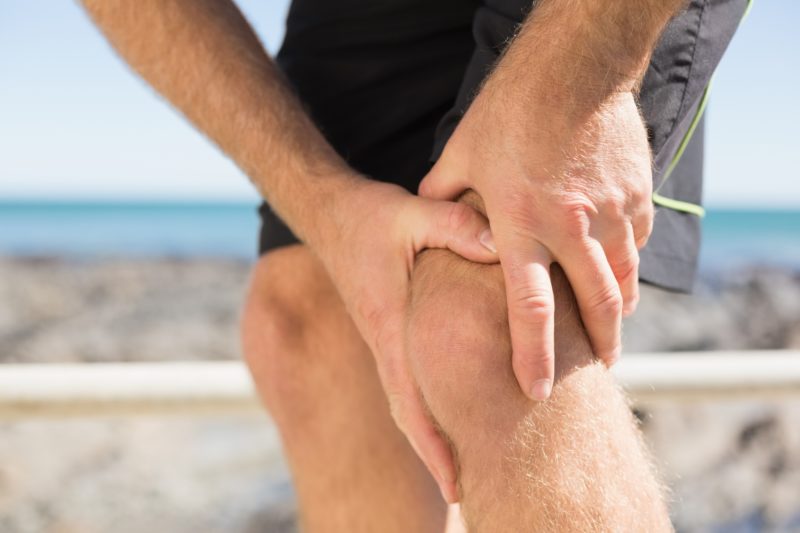
The pain normally presents along the medial (lower) lower 3rd border. The pain is present on running, fast walking or jumping and it can be described as an ache/bruised feeling.
You are able to run/walk through the pain. After exercise the pain usually settles fully with a couple of days. On palpation there is diffuse tenderness along the medial border (highlighted in the picture above). There is no pin point tenderness or night pain.
I commonly see this injury in people who do ‘too much too soon’ meaning a sudden increase in activity level or a change in training programme, this is due to the bone not given adequate time to adapt to the new stress levels. It is important to remember if you have had a couple of months off running you will not be able to return back to the same level straight away, some training is required!
What can you do to treat medial tibial stress syndrome.
There are many ways to treat medial tibial stress syndrome however the evidence shows us there is no one treatment method is better than the other. You will find that the treatment involves more than one treatment option meaning that it is never one treatment the settles the pain.
We know that a lot of people with exercise induced leg pain have gluteal weakness and poor hip function. It is important to remember that when we are assessing leg injuries and foot injuries to consider the function of the chain further up. For example if there is hip weakness/dysfunction we know that the knee internally rotates (moves inwards) and foot pronation will increase.
Activity modification is a key part of the management, this does not mean complete rest, just resting from running or high impact activities, so you can still row, swim use the x-trainer or aqua jog, the Alter G is a very good machine if you have access to one.
We tend to find calf weakness in our patients with medial tibial stress syndrome so commonly we issue heel raise exercises to help strengthen the calf muscle.
There is good evidence to support the use of orthoses (insoles) however, orthoses are like glasses you have different glasses for reading and different glasses are driving, as your eyes are doing different tasks. Sometimes orthoses fail because people have one set that they used to walk and the same set to run in. Again like glasses, if your prescription is wrong, the glasses are no use, orthoses are the same. The fun starts as everyone reacts differently to orthoses, so you cannot apply the same prescription to everyone.
I would like to try and break the myth that orthoses are always long-term, they can be used in the short term if required.
Running re-education can be helpful, increasing your cadence, reducing your stride length, with a midfoot style landing can be helpful.
I would like to stress that it is important to seek professional advice before trying to alter your technique, as again we are all individuals so trying to apply a blanket technique will not work for everyone. Running re-education takes time and dedication.
Tibial Stress Fracture
Stress fracture occurs was a bone has been subjected to excessive amount of force over a period of time, and the bone reaches failure point. I apologise, a bit of science now, however I believe this will make understanding the mechanism of stress fractures easier. Below is a diagram to help explain.
Adapted from (Frost, 1996)
The Utah paradigms above is an adaption of Wolff’s law, which basically states that we need to load bone so that is can remodel, if sufficient force is not put through the bone will become weaker, and if too much force it will result in fracture. This is why people with osteoporosis, it important for them to keep walking. If you are interested in stress and strain, google young’s modulus.
There is some thought that medial tibial stress syndrome is on the same spectrum as a stress fracture, however we are not sure if in untreated medial tibial stress syndrome will result in a stress fracture eventually, if left untreated, though I would not leave it untreated.
How does Tibial stress fracture present?
The presentation is similar to medial tibial stress syndrome, however there are a couple of key differences. With a stress fracture there will be pin point tenderness on palpation. The pain will not cease after activity lasting more than 3 days and there may well be night pain present as well. You do not often see much swelling associated with this injury.
Similarly as medial tibial stress syndrome we see this injury in the too much too soon category and also the overuse category. We know that people with a low calorie diet are risk of a stress fracture.
Also women with an irregular menstrual cycle and low bone density are at risk, combining this with a low calorie diet is something that is known as the female triad. There is worth noting that men can have low calorie diets and be prone to stress fractures.
Officially diagnosed a stress fracture scanning is required. Currently the gold standard is an MRI scan. In the early stages of the stress fracture, x-rays are not sensitive and it can take 2 weeks or more for a stress fracture to show on x-ray. Though a x-ray can rule out a break.
What can you do that treats each Tibial stress fracture?
The treatment depends on severity and location is a stress fracture. Stress fractures which have a low risk of non-union sometimes can be managed with activity modification i.e. reducing your running and doing swimming or cycling until the stress fracture heals. Others may require a period of immobilisation and the timing depends on the location of the stress fracture.
Biomechanical Over load syndrome
Biomechanical over load syndrome is a relatively new term coined by Dr Andrew Franklyn-Miller. This new diagnosis come about after people who had been diagnosed with chronic exertional compartment syndrome, had been getting better with Conservative management. Chronic compartment syndrome was previously diagnosed using intra-compartment pressure testing, however some studies have shown that some people who are pain-free have higher pressures than those who are symptomatic.
It would be wise me to point out that chronic exertional compartment syndrome is still a diagnosis however it is different from acute compartment syndrome which tends after a trauma or surgery and can be a limb threatening pathology.
With regards to the biomechanical overload syndrome we are focusing really on the anterior compartment, the pain occurs due to an overload of the muscles in the anterior compartment of the leg.
Biomechanical overload syndrome would be the 2nd most common cause of exercise induced leg pain I see in clinic. As the term suggests that there is a biomechanical nature to the pain. In a similar fashion to medial tibial stress syndrome there is often weakness within the hips and gluteals. Foot function may well be playing a factor in this, gait analysis is a key way to look at dynamic function whilst running to look at overall lower limb function, as if orthoses are issued, whilst there is hip weakness, it may reduce the effectiveness of the orthoses.
To give you an example following the previous example of the hip weakness and increased foot pronation because of this, this will result in a muscle called tibialis anterior (muscle in the anterior compartment of the leg) working harder to decelerate the velocity (speed) of pronation resulting in overloading and pain within the anterior compartment.
How does biomechanical overload syndrome present?
The pain is described as an intense ache and sometimes a cramp -like sensation, that occurs during running or physical activity. The pain is so severe it causes you to stop their activity and rest, however after up to a short period of rest the pain resolves completely and you are able to return back to running. This cycling keeps repeating itself.
There is often minimal tenderness on palpation along the medial aspect of the tibia. The tibialis anterior muscle can be tender to palpate.
Functional testing we tend to find weakness within the calf muscles and is quite often recreates the pain.
Regards to running style, we tend to see that the people certainly biomechanical overload syndrome tend to be over striders with hip weakness and a heavy heel strike (Heel striking is not bad, again horses for courses).
What can you do to treat biomechanical overload syndrome?
The treatment is similar to medial tibial stress syndrome, there was a particular focus on running technique for biomechanical overload syndrome, a lot of the work by Andrew Franklyn Miller and his colleagues has been focusing on this and is demonstrating good pain reduction. The appropriate treatment which may well be strength and conditioning, stretching, orthoses or footwear advice. With regards to footwear biomechanical overload syndrome we tend to find a shoe with a lower drop (the drop is the difference between the height of the heel and of the forefoot, most ‘normal’ running shoes have around a 10-14mm drop) we are also finding the use of a cushioned shoe or cushioned cover on orthoses helpful, which can help to reduce leg stiffness. Have a read up on Hooke’s law, if you are interested.
Summary
Below is a table of the basic presentations of each of the exercise induced leg pain I discussed, and the likely diagnosis attached to it.
My advice is always should you be experiencing exercise induced leg pain is to get assessed by medical professional, this could be you sports podiatrist, physiotherapist or GP, getting a formal diagnosis made quickly allows the correct treatment plan to be formulated and get you back to running as quickly as possible.
If you have any questions or want to go into anything more detail on one happy to discuss any element of this blog.


